Endocrine System
Endocrine system vs. nervous system
What endocrine glands?
What endocrine glands?
What happens when a target cell recognizes a specific hormone?
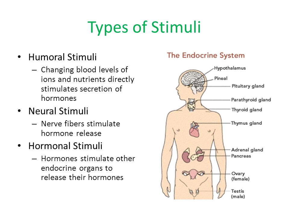
What makes a gland secrete its hormone(s)? Humoral, hormonal and neural stimuli.
Connection between nervous system and endocrine system. Neuroendocrine integration.
Hormonal control of the menstrual cycle.
Hormonal control of the menstrual cycle.
Endocrine system vs. nervous system
Animals, as multicellular organisms, are made of cells, tissues, organs and systems which have to work in a coordinated fashion for the same purposes, one of which is, of course, homeostasis. To achieve this, information has to be exchanged between tissues and organs.
Two coordination systems, namely
i) the nervous system, and
ii) the endocrine system
work together in animals, both vertebrates and invertebrates, to process information from the internal and external environments and produce proper responses.
Those responses controlled by the nervous system are fast, short-term responses. Some slower responses happen over longer periods of time. Growth, development, sexual maturing, sexual cycles, pregnancy, milk secretion in females, metamorphosis in some animals, shedding of skin in others, are examples of such responses.
work together in animals, both vertebrates and invertebrates, to process information from the internal and external environments and produce proper responses.
Those responses controlled by the nervous system are fast, short-term responses. Some slower responses happen over longer periods of time. Growth, development, sexual maturing, sexual cycles, pregnancy, milk secretion in females, metamorphosis in some animals, shedding of skin in others, are examples of such responses.
These slow, long-term processes are under control of chemical messengers called hormones, produced by endocrine (ductless) glands, which release small amounts of these messengers into the bloodstream. Hormones are delivered by blood throughout the organism but they can only be recognised by those organs, tissues or cells that they are specifically targeted at, the target cells, tissues and organs. According to this, hormones act as messengers between distant parts of the organisms in order that these can work in perfect harmony.
Differences between the nervous system and the endocrine system:


What are endocrine glands?
A gland is an organ, sometimes just a tissue, the function of which is to secrete substances. Glands and glandular tissues exist both in animals and plants, and contain secretory cells that are specialized in producing and releasing a specific substance with a specific function. In animals, glands are classified into two groups according to the substances they produce and the way these substances are released:
- Exocrine glands produce a variety of substances other than hormones, and release these substances through ducts or tubes onto epithelial surfaces like the skin or mucous epithelia like those lining the gastrointestinal, urogenital and respiratory tracts. Examples of exocrine glands and cells are the salivary glands, the sweat glands, the fat glands, the tear glands, the mammary glands in females, the chief, oxintic and goblet secretory cells in the stomach, the exocrine pancreas, the prostate, Cowper´s glands and seminal vesicles in males, and many others.
- Endocrine glands produce hormones, chemical messengers that are poured directly into the bloodstream rather than through a duct, hence the name ductless glands to refer to endocrine glands. These are the glands that we are going to focus our attention on.
The picture below shows the location of some important endocrine glands, the hormones produced by which and their functions are explained in the table that follows. The hypothalamus and the pituitary gland deserve to be explained separately, as they constitute the connection between the nervous and endocrine system.

What happens when a target cell recognises a specific hormone?
One given target cell in a target organ or tissue has receptor proteins which recognise and bind to specific hormones. These receptors can be located on the surface of the cell, that is, can be membrane proteins, or be present in the cytoplasm of the cell. Recognition of a hormone by a cell is followed by a variety of responses which depend on the type of target cell and the hormone, for instance
- contraction of muscle fibres, as is the case of contraction of the uterus during delivery,
- Synthesis and secretion of secretory products by exocrine glands,
- breakdown of storage molecules like glycogen, which is broken down into glucose molecules, of fats, which are broken down into fatty acids.
- Synthesis and secretion of hormones by endocrine glands. Some endocrine glands secrete their hormones as a response to recognition of a hormone seceted by another gland.
What makes a gland secrete its hormone(s)?
The same as the nervous system makes responses to stimuli collected by receptors, glands need stimulation to produce and secrete their hormones. Three types of stimuli for glands have been described, namely humoral, hormonal and neural.
- Humoral stimuli. The gland detects a high (or low) concentation of a specific chemical in blood (more accurately in the interstitial fluid) and responds by secreting a hormone the effect of which is to decrease (or increase) the level of this substance in blood.
Example 1: Insulin and glucagon are produced by the endocrine pancreas as a response to changes in concentration of glucose in blood (humoral stimulus).
After a meal, digestion and absorption result in high levels of glucose and amino acids in blood. This increase in blood glucose (and amino acids) is detected by the 𝞫-cells in the islets of Langerhans in the pancreas, which triggers insulin secretion by these cells. Insulin reaches its target organs and tissues, namely liver, muscles and adipose tissue, via blood, and stimulates uptake of glucose by these organs and tissues, and conversion to glycogen in muscles and liver, or fats in the adipose tissue.
 A portion of the pancreas as seen under a light microscope. The lighter cells in the middle of the picture make up an islet of Langerhans, which contains endocrine secretory cells, namely 𝞫-cells, responsible for secreting the hormone insulin as a response to high concentration of glucose in blood, and 𝞪-cells, responsible for secreting the hormone glucagon as a response to low concentration of glucose in blood. The darker cells around the islet of Langerhans are exocrine secretory cells, which secrete the digestive enzymes present in the pancreatic juice as a reponse to recognition of the hormone secretin, produced by the duodenum an jejunum. Secretin is produced by the small intestine as a response to acidity provided by the hydrochloric acid in the chyme which comes from the stomach (humoral stimulus).
A portion of the pancreas as seen under a light microscope. The lighter cells in the middle of the picture make up an islet of Langerhans, which contains endocrine secretory cells, namely 𝞫-cells, responsible for secreting the hormone insulin as a response to high concentration of glucose in blood, and 𝞪-cells, responsible for secreting the hormone glucagon as a response to low concentration of glucose in blood. The darker cells around the islet of Langerhans are exocrine secretory cells, which secrete the digestive enzymes present in the pancreatic juice as a reponse to recognition of the hormone secretin, produced by the duodenum an jejunum. Secretin is produced by the small intestine as a response to acidity provided by the hydrochloric acid in the chyme which comes from the stomach (humoral stimulus).
Conversely, after a period of fasting or during intense physical activity the level of glucose in blood decreases, which is detected by the 𝞪-cells that share the islets of Langerhans with the 𝞫-cells. The 𝞪-cells respond by secreting the hormone glucagon, which reaches its target organs, liver and muscles, via blood. Once there glucagon promotes breakdown of glycogen back into glucose, which returns to blood to increase blood glucose level. This is a very good example of the contribution of the endocrine systen to homeostasis.
Example 2: The parathyroid glands respond to low level of blood calcium.
 Calcium is a metallic chemical element which serves vital functions in the organism, notably muscle contraction. Low levels of blood calcium stimulate the C-cells in the parathyroid glands to produce a hormone, the parathyroid hormone PTH, the effects of which are release of calcium from the bones and enhanced reabsorption of calcium by renal tubules, thereby increasing calcium in blood to normal levels.
On the contrary, when the concentration of calcium in blood is too high, C-cells located in the thyroid gland respond by secreting calcitonin, a hormone whose target organs are bones and kidneys, too. Calcitonin in bones inhibits breakdown of calcium phosphate and release of calcium into the blood. In kidneys calcitonin inhibits selective reabsorption of calcium by the renal tubules, excess calcium being excreted with urine.
The parathyroid hormone PTH, secreted by the parathyroid gland, and calcitonin, secreted by the thyroid gland, have antagonistic effects to one another.
 Calcium is a metallic chemical element which serves vital functions in the organism, notably muscle contraction. Low levels of blood calcium stimulate the C-cells in the parathyroid glands to produce a hormone, the parathyroid hormone PTH, the effects of which are release of calcium from the bones and enhanced reabsorption of calcium by renal tubules, thereby increasing calcium in blood to normal levels.
On the contrary, when the concentration of calcium in blood is too high, C-cells located in the thyroid gland respond by secreting calcitonin, a hormone whose target organs are bones and kidneys, too. Calcitonin in bones inhibits breakdown of calcium phosphate and release of calcium into the blood. In kidneys calcitonin inhibits selective reabsorption of calcium by the renal tubules, excess calcium being excreted with urine.
The parathyroid hormone PTH, secreted by the parathyroid gland, and calcitonin, secreted by the thyroid gland, have antagonistic effects to one another.
- Hormonal stimuli. Some glands are stimulated to secrete their hormones by hormones secreted by other glands referred to as tropic hormones. In other words, tropic hormones are hormones whose target organs are endocrine glands. Most tropic hormones are produced by a small yet vital gland, the size of a pea, located at the base of the brain, known as the pituitary gland or hypophysis, also referred to as the master gland, as its hormones control secretion by other endocrine glands which, in turn, control many body functions.
Example 3: Testicles and ovaries are stimulated by hormones from the pituitary gland to produce male and female sex hormones (hormonal stimulus).
One hormone produced by the pituitary gland named luteinizing hormone, LH, stimulates production of sexual hormones by the testicles called androgens, the most important of which is testosterone, resposible for those sexual characteristics which make males different from females (distribution of body hair and fat, larger muscles, sexual behaviour, for instance). In females, LH estimulates secretion of another hormone, progesterone, by the ovaries. Progesterone is responsible for the development of the internal mucous lining of the uterus, the endometrium, during the the first two weeks of the menstrual cycle, between menstruation and ovulation. The function of the endometrium is to provide the embrio with shelter and support in case of pregnancy. If fertilization of the egg cell does not occur most of the endometrium will be shed during menstruation.
- Neural stimuli. Besides the hypothalamus, which will be dealt with later, some glands are under direct control of the nervous system, more accurately, under control of the sympathetic division of the autonomic nervous system. That is the case of the adrenal glands.
Example 4: The adrenal medulla secretes adrenaline as a result of neural stimulation by the sympathetic division of the peripheric nervous system.


The adrenal glands on top of the kidneys.
The adrenal glands or suprarenal glands are paired glands located on top of either kidney. They consist of an outer adrenal cortex around an inner adrenal medulla, both with endocrine functions. The adrenal medulla secretes the hormone adrenaline (or adrenalin, epinephrine in America) under stimulation of nerve fibres (axons) from the sympathetic division of the autonomic nervous system (running along the greater thoracic splanchnic nerve) as a response to stressing, physically demanding or dangerous situations. In fact, secretion of adrenaline by the adrenal medulla has similar effects on the same organs and tissues as the stimulation by the sympathetic division of the autonomic nervous system: breakdown of glycogen in muscles and liver, and fats in adipose tissue, increase in rate and power of heart beat, dilation of brochi and bronchioles, inhibition of digestion and excretion, among others. Unlike other hormones, the effects of adrenaline are fast and short-lived.
Connection between nervous system and endocrine system. Neuroendocrine integration.
The nervous and endocrine systems are not independent of each other, they exchange information in such a way that nervous impulses can be translated into chemical information in the neuroendocrine organs. These organs, the most important of which are the hypothalamus and the hypophysis or pituitary gland, are made of neurosecretory cells, that is, neurons which produce chemical messengers called neurohormones, that are poured into the bloodstream as a response to nerve impulses reaching these cells, which play the same role as endocrine secretory cells. To the question: What is there at the end of one axon of a neuron? there ae three possible answers:
- A dendrite of a post-synaptic neuron, the connection between both neurons being named a synapse.
- An effector, at the end of efferent (motor) axons.
- A blood capillary.

If the answer is number 3 the situation is like that depicted in the diagram on the right. At the end of the axon of this neuron there is neither another neuron nor an effector, but a blood capillary. The chemical messengers secreted by the neuron are poured into the bloodstream, which carries them to their specific target organs as is the case with hormones produced by endocrine glands.
Are these chemical messengers neurotransmitters, because they are produced by a neuron, or are they hormones, because they are transported by blood? They are referred to as neurohormones produced by neuroendocrine cells, responsible for neuroendocrine integration.
Are these chemical messengers neurotransmitters, because they are produced by a neuron, or are they hormones, because they are transported by blood? They are referred to as neurohormones produced by neuroendocrine cells, responsible for neuroendocrine integration.
 The most important sites of neuroendocrine integration are the hypothalamus and the hypophysis or pituitary gland (master gland). They are physically connected by a narrow pituitary stalk or infundibulum, and jointly make what is referred to as the hypothalamic-hypophyseal axis.
The pituitary gland is divided into two parts, namely
- The anterior hypophysis or adenohypophysis , and
- the posterior hypophysis or neurohypophysis, which can be considered as an extension of the hypothalamus.
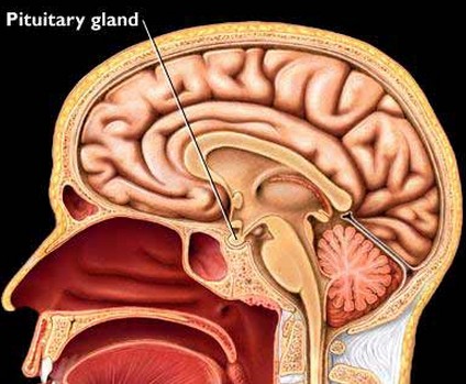
The hypothalamus and the pituitary gland or hypophysis, connected by the pituitary stalk or infundibulum, at the base of the cerebrum.


The hypothalamus is the neurosecretory region of the brain. As a part of the brain is made of neurons, but these neurons pour the chemical messengers they produce into blood capillaries. We call these neurons neuroendocrine cells and the substances they produce, neurohormones, which means hormones produced by neurons. There are two types of neurons in the hypothalamus:

- Neurons with long axons which run along the pituitary stalk and reach the posterior pituitary (neurohypophysis). These neurones secrete neurohormones that are poured into capillary beds through which these neurohormones, oxytocin and antidiuretic hormone ADH, reach their target organs, namely mammary glands, uterus (oxytocin) and kidneys (ADH), via the bloodstream.(Example 5)
- Neurons whith shorter axons which discharge their neurohormones in the hypophyseal portal system, which takes these neurohormones to the anterior pituitary (adenohypophysis). These neurohormones stimulate (or inhibit) the endocrine secretory cells in the anterior pituitary to secrete its hormones. (Examples 6 and 7).

The anterior pituitary or adenohypophysis secretes a number of hormones under stimulation from releasing hormones or releasing factors (hormonal stimulus) secreted by neurosecretory cells in the hypothalamus. These releasing hormones are discharged into the blood vessels of the hypothalamus-hypophyseal portal system through which they travel the short distance to the adenohypophysis. Once there each type of releasing hormone brings about secretion of a specific hormone, some of which are tropic hormones:

Light microscope picture of an ovary showing graafian follicles about to release their egg cells, a process referred to as ovulation.
In these animals, during copulation, afferent fibres from the genital organs inform the brain about sexual intercourse taking place. This information reaches neurosecretory cells in the hypothalamus that are stimulated to secrete a releasing hormone, the LHRH (luteinizing hormone-releasing hormone). LHRH is released into the hypothalamus-hypophyseal portal system located in the pituitary stalk, to eventually reach the anterior hypophysis, where this releasing hormone is recognised by specific endocrine secretory cells. Recognition results in these secretory cells releasing luteinizing hormone LH into the bloodstream (hormonal stimulus). LH has different target cells in different locations, both in males anf females, with different effects, one of which is to trigger release of the egg cell by a graafian follicle, in other words, ovulation.
Thyroxine secretion by the thyroid gland is under hormonal control by a tropic hormone from the adenohypophysis or anterior pituitary gland, this hormone being named thyroid-stimulating hormone TSH. TSH secretion by the adenohypophysis is, in turn, stimulated by a releasing hormone from the hypothalamus named TRH (thyrotrophin-releasing hormone). Information from receptors about internal and external, physical or chemical environmental conditions reach the brain. The hypothalamus is a part of the brain and becomes aware of these conditions, and will make a proper response if these conditions are a threat to homeostasis. For instance, how does the organism respond to exposure to cold?

Maintenance of body temperature upon exposure to cold by the hypothalamic-pituitary-thyroid axis, involving one neurohormone (TRH) and two hormones (TSH and thyroxine)

A good example of neurohormonal control by the hypothalamus and the hypophysis is the control of the menstrual cycle.
The anterior hypophysis secretes two hormones, namely luteinizing hormone LH and follicle stimulating hormone FSH, under stimulation by releasing hormones (releasing factors) from the hypothalamus.


Oestrogens and progesterone, produced by the corpus luteum under stimulation of LH, are responsible for thickening and maintenance of the endrometrium, in preparation for implantation in case fertilisation happened.
Gland
Exocrine gland
Endocrine gland
Hormone
Target cell, tissue of organ
Humoral stimulus
Hormonal stimulus
Neural stimulus
Islets of Langerhans
Insulin
Glucagon
Parathyroid gland
Thyroid gland
Adrenal medulla
Adrenalin
Neuro endocrine organs
Hypothalamus
Hypophysis or Pituitary gland
Neurohormones
Neuro endocrine cells
Anterior Hypophysis
Posterior Hypophysis
Hypophyseal portal system
Pituitary stalk
Tropic hormones
Releasing hormones
- Hormones secreted by the adenohypophysis include growth hormone (somatotrophin) and prolactin. Growth hormone acts on several target organs and tissues. It stimulates protein synthesis and breakdown of fats as a source of energy, as well as promoting growth of long bones in arms and legs. Prolactin for its part stimulates (along with oxytocin) milk secretion by mammary glands, among other functions.
- Tropic hormones are those hormones whose target organs are other endocrine glands, notably the thyroid gland and the gonads, that is, ovaries and testicles.
Most endocrine glands are one way or another, directly or indirectly, under control of hormones from the pituitary gland, which amounts to saying that most endocrine glands are under control of the hypothalamus, that is, the brain.
Example 5: Milk secretion by
females is under control of the hypothalamic-hypophyseal axis. Not all women
breastfeed their newborn babies. The fact that a woman has given birth does not
mean that her mammary glands (exocrine) will produce milk. For this to
happen hormonal stimulation is required. When the baby suckles, the nipples act
as receptors and afferent fibres inform the brain about this action taking
place. This information from the nipples reaches neurons in the hypothalamus, the axons of which run through the pituitary stalk down to the posterior
hypophysis or neurohypophysis. These neurosecretory cells produce a
neurohormone called oxytocin, which is stored and eventually discharged into
capillary beds inside the neurohypophysis. From here oxytocin enters the
bloodstream to be recognised by its target organ, the mammary glands, where it
stimulates milk production.
A hormone from the anterior hypophysis, prolactin, has similar effects on the mammary glands as oxytocin, its secretion being stimulated by suckling likewise.
A hormone from the anterior hypophysis, prolactin, has similar effects on the mammary glands as oxytocin, its secretion being stimulated by suckling likewise.
The neurohypophysis or posterior hypophysis can be considered as an
extension of the hypothalamus rather than a gland. It houses the terminals of
axons of neurosecretory cells from the hypothalamus. Neurohormones from these
neurons, like oxytocin, are stored here before entering the blood circulation.
Oxytocin is also involved in uterine smooth muscle contraction during
childbirth. When women are in labour, a word used to refer to the process of giving birth, they are usually given an injection of oxytocin, a hormone which prompts contraction by the uterine smooth muscle, the myometrium.
Example 6: Some mammals
like cats and rabbits are induced ovulators. Ovulation in human females is
expected to happen every 28 days and is thought to be under control of an
"internal clock" located somewhere inside the brain. In contrast, the
females of other mammals, like cats and rabbits are known to ovulate after, or
as a result of copulation, hence the name induced ovulators.

Light microscope picture of an ovary showing graafian follicles about to release their egg cells, a process referred to as ovulation.
In these animals, during copulation, afferent fibres from the genital organs inform the brain about sexual intercourse taking place. This information reaches neurosecretory cells in the hypothalamus that are stimulated to secrete a releasing hormone, the LHRH (luteinizing hormone-releasing hormone). LHRH is released into the hypothalamus-hypophyseal portal system located in the pituitary stalk, to eventually reach the anterior hypophysis, where this releasing hormone is recognised by specific endocrine secretory cells. Recognition results in these secretory cells releasing luteinizing hormone LH into the bloodstream (hormonal stimulus). LH has different target cells in different locations, both in males anf females, with different effects, one of which is to trigger release of the egg cell by a graafian follicle, in other words, ovulation.
Example 7. The thyroid gland is under hypothalamic control. The thyroid gland is a
small (≈ 5 cm. across) butterfly-shaped endocrine gland located at the front of
the neck, just below the Adam´s apple.
The thyroid gland
produces three different hormones, the most important, of which is iodine-containig thyroxine,
T4. There are multiple target organs and tissues for thyroxine (or thyroxin),
the general effect of which is to increase the energy metabolism in tissues,
which results in
- enhanced thermogenesis,
- increased basal metabolic rate,
- increased cell respiration (mitochondria) and, as a consequence, increased oxygen consumption,
- enhanced glycogenolysis in muscle and liver,
- enhanced breakdown of fats in adipose tissue,
- enhanced absorption of glucose in gut and uptake of glucose by cells,
- Cold environmental conditions are noticed by receptors in our skin.
- Information reaches the brain through afferent fibres.
- Neurosecretory cells in the hypothalamus, a part of the brain, synthesise a releasing hormone (releasing factor) named TRH.
- TRH is released in the hypophyseal portal system (located in the pituitary stalk), and conveyed to the anterior pituitary by blood in the portal system vessels.
- THR is recognized by secretory cells in the anterior pituitary responsible for secretion on TSH.
- TSH, a tropic hormone, is secreted and poured into the bloodstream.
- TSH in blood is recognized by the thyroid gland, which is stimulated to produce thyroxine.
- Thyroxine is poured into the bloodstream and reaches its various target organs.
- Increased energy metabolism results in generation of heat.

Maintenance of body temperature upon exposure to cold by the hypothalamic-pituitary-thyroid axis, involving one neurohormone (TRH) and two hormones (TSH and thyroxine)

Hormonal control of the menstrual cycle
A good example of neurohormonal control by the hypothalamus and the hypophysis is the control of the menstrual cycle.
The anterior hypophysis secretes two hormones, namely luteinizing hormone LH and follicle stimulating hormone FSH, under stimulation by releasing hormones (releasing factors) from the hypothalamus.
- FSH stimulates ripening of graafian follicles inside the ovary. Each egg cell is kept inside a graafian follicle until ovulation.
- LH triggers ovulation and formation of the corpus luteum, a secretory tissue which develops inside the empty graaafian follicle. The function of the corpus luteum is to secrete other hormones, namely progesterone and oestrogens.


A graafian follicle about to relase its ovum (left), and the corpus luteum that develops inside an empty graafian follicle (right).

Ovaries, fallopian tubes and womb
The menstrual cycle can be conveniently divided into an ovarian cycle, all the things that happen in the ovary over a menstrual cycle, and the uterine cycle, all the things that happen in the uterus over a cycle. An average menstrual cycle takes 28 days, but it can be shorter or longer.


Ovaries, fallopian tubes and womb
The menstrual cycle can be conveniently divided into an ovarian cycle, all the things that happen in the ovary over a menstrual cycle, and the uterine cycle, all the things that happen in the uterus over a cycle. An average menstrual cycle takes 28 days, but it can be shorter or longer.


Uterine cycle Ovarian cycle
Consider the day that menstruation comes as the first day of the cycle. During the first 3-7 days menstruation takes place. The endometrium degenerates and is expelled. The endometrium is the mucous internal lininig of the uterus, rich in blood vessels, which provides fertilised ova with protection and nourishment in case of fertilisation. During the rest of the cycle the endometrium will grow and thicken again in preparation for a a pregnancy. In the absence of fertilisation, the new endometrium will be shed.
Meanwhile in the ovaries, normally one of them, a graafian follicle develops during the first two weeks of the cycle, under stimulation by FSH (follicle stimulating hormone). Maturation of the follicle is completed around the fourteenth day of the cycle and is followed by release of the ovum inside, ready for fertilisation, that is, ovulation, under stimulation by LH (luteinizing hormone). The ovum will travel the short way to the uterus via the fallopian tube, where fertilisation may happen or not.
Consider the day that menstruation comes as the first day of the cycle. During the first 3-7 days menstruation takes place. The endometrium degenerates and is expelled. The endometrium is the mucous internal lininig of the uterus, rich in blood vessels, which provides fertilised ova with protection and nourishment in case of fertilisation. During the rest of the cycle the endometrium will grow and thicken again in preparation for a a pregnancy. In the absence of fertilisation, the new endometrium will be shed.
Meanwhile in the ovaries, normally one of them, a graafian follicle develops during the first two weeks of the cycle, under stimulation by FSH (follicle stimulating hormone). Maturation of the follicle is completed around the fourteenth day of the cycle and is followed by release of the ovum inside, ready for fertilisation, that is, ovulation, under stimulation by LH (luteinizing hormone). The ovum will travel the short way to the uterus via the fallopian tube, where fertilisation may happen or not.
If fertilisation does nor occur
The corpus luteum starts to develop inside the empty graafian follicle under stimulation by FSH, but in the absence of fertilisation it degenerates by the end of the cycle. The endometrium reaches its maximum thickness. At the end of the cycle it degenerates an is shed: menstruation comes.
Fertilisation commonly happens while the egg cell is in the fallopian tube, on its way to the uterus. Upon reaching the uterus, the fertilised egg becomes embedded in the mucous endometrium, a process referred to as implantation. Inside the ovary the corpus luteum does not degenerate and becomes an endocrine tissue, responsible for producing progesterone and oestrogens, the most important of which is oestradiol. Both hormones play several roles during pregnancy, one of which is to prevent further ovulations. For this reason progesterone and oestrogens are used in contraceptive pills.
If there is fertilisation
Oestrogens and progesterone, produced by the corpus luteum under stimulation of LH, are responsible for thickening and maintenance of the endrometrium, in preparation for implantation in case fertilisation happened.
VOCABULARY
Gland
Exocrine gland
Endocrine gland
Hormone
Target cell, tissue of organ
Humoral stimulus
Hormonal stimulus
Neural stimulus
Islets of Langerhans
Insulin
Glucagon
Parathyroid gland
Thyroid gland
Adrenal medulla
Adrenalin
Neuro endocrine organs
Hypothalamus
Hypophysis or Pituitary gland
Neurohormones
Neuro endocrine cells
Anterior Hypophysis
Posterior Hypophysis
Hypophyseal portal system
Pituitary stalk
Tropic hormones
Releasing hormones
Gland
|
Hormone
|
Target organ/tissue
|
Response
|
Thyroid
|
Thyroxine
| ||
Parathyroid glands (x4)
| |||
Adrenal glands (x2)
| |||
Pancreas
| |||
Ovaries (x2)
| |||
Testicles (x2)
| |||
Thymus
|




0 Comments